Home
/ Animal Cell Diagram Golgi Body - Animal Cells Animal Cell Cell Model Cell Biology : The golgi adds small molecules (that act as stamps) onto lipids, carbohydrates and proteins, packages them up.
Animal Cell Diagram Golgi Body - Animal Cells Animal Cell Cell Model Cell Biology : The golgi adds small molecules (that act as stamps) onto lipids, carbohydrates and proteins, packages them up.
Animal Cell Diagram Golgi Body - Animal Cells Animal Cell Cell Model Cell Biology : The golgi adds small molecules (that act as stamps) onto lipids, carbohydrates and proteins, packages them up.. The golgi body is in a membrane bound form (stack of membrane bound vesicles) which is essential for packing and transporting macromolecules. The golgi bodies store the different chemicals made by different parts of the cells until they are ready to be released into the cytoplasm. That cells can be of different shapes and sizes. Color the remaining space in the cell, which is called cytosol or cytoplasm, a liquid in which the organelles live. It is referred to as the manufacturing and the shipping center of the cell.
Complete with videos, quizzes, links and summary tables. The cell membrane, or plasma membrane, is a golgi apparatus: The golgi adds small molecules (that act as stamps) onto lipids, carbohydrates and proteins, packages them up. Organelles are labelled as follows Lysosomes are produced by the golgi apparatus and appear like small spherical bodies featuring a single.
Animal Cell Diagram 3d Warehouse from 3dwarehouse.sketchup.com The role and function of the plasma membrane; Cell membrane nucleolus golgi body mitochondrion. The golgi body, sometimes referred to as either the golgi apparatus or golgi complex, is responsible for the packing of proteins into vesicles. The cell membrane, or plasma membrane, is a golgi apparatus: Plant cell drawing animal cell drawing human cell diagram plant cell diagram biology animal cells have a single highly complex and prominent golgi apparatus. Each centriole is a ring of nine groups of fused microtubules. Golgi bodies create hormones from proteins. Tem micrograph of golgi body, visible as a stack of semicircular black rings near the bottom.
A comparison of plant and animal cells using labelled diagrams and descriptive explanations.
Color the remaining space in the cell, which is called cytosol or cytoplasm, a liquid in which the organelles live. A golgi body, the golgi complex or the golgi apparatus. Organelles are labelled as follows It has been estimated that humans detailed diagram of lipid bilayer cell membrane. Golgi apparatus (or golgi bodies). Cytoplasm, ribosomes, rough endoplasmic reticulum; Smooth er nucleus free ribosome vacuole. Mitochondria, endoplasmic reticulum, golgi apparatus, lysosomes, diagram of a typical animal cell. Golgi body (golgi apparatus/golgi complex). Diagram showing golgi bodies found in animal cells. Tem micrograph of golgi body, visible as a stack of semicircular black rings near the bottom. Draw the golgi body inside the cell. Smooth endoplasmic reticulum, mitochondria, golgi bodies, lysosomes.
Lysosomes are produced by the golgi apparatus and appear like small spherical bodies featuring a single. Each organelle has a different purpose inside the cell. A comparison of plant and animal cells using labelled diagrams and descriptive explanations. The golgi bodies store the different chemicals made by different parts of the cells until they are ready to be released into the cytoplasm. The golgi body was discovered by the italian physician camillo golgi.
Animal Cell Anatomy Enchanted Learning from www.enchantedlearning.com A golgi body, the golgi complex or the golgi apparatus. Golgi bodies process and transfer substances to the correct location. An animal cell diagram is a great way to learn and understand the many functions of an animal cell. After completing this section, you should know: Draw the golgi body inside the cell. The golgi body packages and sends complex molecules around and out of the cell. Animals are made up of basic building blocks called the animal cell. Each centriole is a ring of nine groups of fused microtubules.
The golgi body is in a membrane bound form (stack of membrane bound vesicles) which is essential for packing and transporting macromolecules.
5th grade science and biology. Let us look at animal cell parts and functions, using diagrams and illustrations. Printable animal cell diagram to help you learn the organelles in an animal cell in preparation for your test or quiz. It has been estimated that humans detailed diagram of lipid bilayer cell membrane. Lysosomes are produced by the golgi apparatus and appear like small spherical bodies featuring a single. Golgi apparatus (or golgi bodies). Smooth er nucleus free ribosome vacuole. The golgi body, sometimes referred to as either the golgi apparatus or golgi complex, is responsible for the packing of proteins into vesicles. The golgi apparatus is made up of stacks of membranous layers that are referred to as golgi bodies. The golgi bodies store the different chemicals made by different parts of the cells until they are ready to be released into the cytoplasm. The golgi body was discovered by the italian physician camillo golgi. There are over 200 different cell types in the human body, each with a very specific job. Cytoplasm, ribosomes, rough endoplasmic reticulum;
It has been estimated that humans detailed diagram of lipid bilayer cell membrane. Diagram of animal cell, created with biorender.com. Organelles are labelled as follows Kidneys are a pair of excretory organs located in lower back of our body.each kidney is reddish. A comparison of plant and animal cells using labelled diagrams and descriptive explanations.
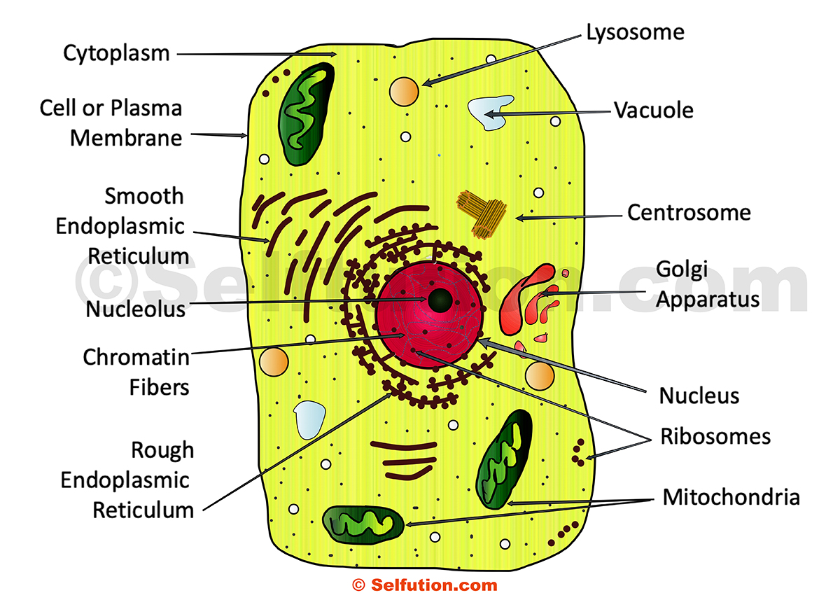
Structure And Function Of A Cell And Its Organelles Selftution from selftution.com Golgi apparatus (or golgi bodies). The golgi apparatus is made up of stacks of membranous layers that are referred to as golgi bodies. Take a visual tour of the animal cell. Complete with videos, quizzes, links and summary tables. Animal cell anatomy diagram structure with all parts nucleus smooth rough endoplasmic reticulum cytoplasm golgi apparatus. Organelles are labelled as follows A golgi body, the golgi complex or the golgi apparatus. Smooth endoplasmic reticulum, mitochondria, golgi bodies, lysosomes.
Draw the golgi body inside the cell.
Animal cells are typical of the eukaryotic cell, enclosed by a plasma membrane and containing a the lack of a rigid cell wall allowed animals to develop a greater diversity of cell types, tissues, and the nuclei are stained with a red probe, while the golgi apparatus and microfilament actin network. The golgi body is in a membrane bound form (stack of membrane bound vesicles) which is essential for packing and transporting macromolecules. Golgi body (golgi apparatus/golgi complex). Animal cell anatomy diagram structure with all parts nucleus smooth rough endoplasmic reticulum cytoplasm golgi apparatus. Every animal cell has two of these small organelles (made of microtubules) and they help organize cell division (like a teaching assistant who help out near the office). Take a visual tour of the animal cell. Let`s draw a typical animal cell. Lysosomes are produced by the golgi apparatus and appear like small spherical bodies featuring a single. Diagram of animal cell, created with biorender.com. Cytoplasm, ribosomes, rough endoplasmic reticulum; Animal cells differ from plant cells in several regards though, including the lack of vacuoles, chloroplasts, and cell walls. Animal cells, unlike plant and fungi cells, do not have a cell wall. Draw the golgi body inside the cell.
Share :
Post a Comment
for "Animal Cell Diagram Golgi Body - Animal Cells Animal Cell Cell Model Cell Biology : The golgi adds small molecules (that act as stamps) onto lipids, carbohydrates and proteins, packages them up."


Post a Comment for "Animal Cell Diagram Golgi Body - Animal Cells Animal Cell Cell Model Cell Biology : The golgi adds small molecules (that act as stamps) onto lipids, carbohydrates and proteins, packages them up."